Online scheduling is available for our Stamford, CT location ONLY
Stamford Scheduling (203) 324-5719 8:00 AM – 4:00 PM, weekdays
Garden City Scheduling (516) 227-3377 9:00 AM – 5:00 PM, weekdays
Learn about our mobile scanning options at www.dermvan.com
Melanoscan …..bringing 21st century technology to the fight against deadly melanoma.
Are you at risk? Complete our Skin Cancer Risk Assessment!
The Melanoscan Whole-body digital imaging system was developed by one of the country’s leading academic scientists.
Creating your Melanoscan Whole-body digital Photographic Image couldn’t be easier: it’s a completely safe procedure, involving no radiation, prior preparation or discomfort. The entire scanning process takes only a few minutes, and occurs in a comfortable, private environment.
Melanoscan was created to capture total body images which can then be reviewed and interpreted by a dermatologist. Sequential scanning over time makes it easier for both the patient and physician to identify subtle skin and mole changes.
Using this state of the art new technology, the critical initial signs of dangerous melanoma can be detected at an earlier stage of growth and easily treated.
Melanoma Revealed through Melanoscan Sequential Photography
The Power of Time Lapse Change Detection:
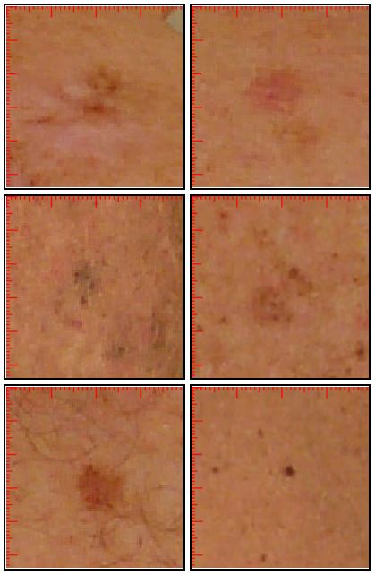
Would you have spotted the change in each lesion signifying Melanoma without the time lapse image comparison?
• Melanoma may be recognized earlier by lesion image comparison, utilizing time lapse scanning.
• Melanomas detected early, before they have a chance to grow in size and thickness (depth) are almost always curable.
• However, melanomas left unchecked and undetected, are potentially fatal.
Without sequential scanning, early melanomas are easily missed by patients and dermatologists alike, but with the Melanoscan technology, your spots of concern can be identified, examined by your dermatologist, tracked and monitored over time.
If any of the above apply to you, consider a small investment of time and money that could potentially save your life.
And please remember:
Melanoma can and does strike people with dark skin, hair and eyes, even if their risk profile is considered low.
Take the FREE Online Melanoma Risk Assessment survey.
If you scored 10 or higher on our Skin Cancer Risk Assessment survey, contact us today at (203) 324-5719 to schedule an appointment at our Stamford, CT clinic
or
(516) 227-3377 to schedule an appointment at our Garden City, NY clinic, and take advantage of this new and potentially life saving Melanoscan technology.
If you are in the moderate or high risk for developing melanoma or other skin cancers, we urge you to schedule an appointment for a Melanoscan. After completing your Melanoscan, it should be reviewed (preferably within a few weeks), by a dermatologist who will also perform a full body skin exam focusing on any spots identified in your images as well as any other lesions of concern.
Pamela Basuk, MD
2011 Union Boulevard Suite 1
Bay Shore, NY 11706
631-666-2900
www.basukdermatology.com
Theodore Daly, MD
901 Stewart Avenue, Suite 205
Garden City, NY 11530
516-227-3377
www.gardencityderm.com
Rhett Drugge, MD
50 Glenbrook Road, Unit 1C
Stamford, CT 06902
203-324-5719
www.thebestdermatologist.com
Rena Fortier, MD
1051 Long Ridge Road
Stamford, CT 06903
203-329-7960
www.longridgedermatology.com
Antoinette Notaro, MD
13405 Main Road
Mattituck, New York 11952
631-298-1122
www.antoinettenotaromd.com
Erin Schoor, MD
180 East Pulaski Road
Huntington Station, NY 11746
631-425-2121
www.hmgpc.com
Erin Walker, MD
210 Westchester Avenue
White Plains, NY 10604
914-682-6426
www.westmedgroup.com
Saryna Young, MD
210 Westchester Avenue
White Plains, NY 10604
914-682-6426
www.westmedgroup.com
Donald Savitz, M.D. – Long Ridge Dermatology, Stamford
Ellen Nairdorf, M.D. – Stamford
Rebecca Hall, M.D. – Stamford
Rand Werbitt, M.D. – Stamford
Samuel Goettler, M.D. – Stamford
Fern Meyer, M.D. – Stamford
Make an appointment by contacting the Dermatology Imaging Center at (203) 324-5719 or by e-mailing us at TheBestDermatologist@gmail.com.
You may also schedule an appointment by sending us a
The Melanoscan imaging centers are located at:
50 Glenbrook Road Unit 1C
Stamford, CT 06902
Fax: (203) 323-7485
Stamford is 45 minutes by train from New York’s Grand Central Terminal. The office is a 15 minute walk from the Metro North/Stamford train station, and taxis are available.
And:
901 Steward Avenue Suite 205
Garden City, NY 11530
Call (516) 227-3377 to schedule an appointment
PLEASE NOTE: Online scheduling in not yet available for our Garden City location
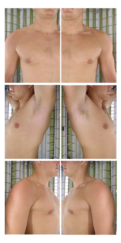
The patented Melanoscan® digital imaging system enables dermatologists to identify changes in skin and moles by comparing photographic maps of the entire body taken at different times. For example, a patient who is at high risk for skin cancer may take pictures to be compared every six months. In this way, physicians can potentially identify small changes in skin topography that may indicate melanoma. Lesions that are unchanged over time are seldom malignant.
This advanced skin cancer monitoring system is a combination of three choreographed positioning techniques and hi-tech software algorithms to enhance the reproducibility of the photos.
After entering the scanning booth, the patient assumes the following position:
Once the front and back scans have been completed, the patient may exit the booth, get dressed and return to the receptionist. The whole process takes only a few minutes.
The patented Melanoscan® system is a state-of-the-art, fully automated, 25 camera whole body digital scanner. It is currently operational in the U.S. in Connecticut and New York states, having been in steady use and rigorously tested for more than a decade.
Nationwide availability is planned for 2019.
The lead inventor of the Melanoscan imaging system is Dr. Rhett Drugge, a board-certified dermatologist currently practicing in Stamford, CT. A graduate of Harvard University, Dr. Drugge received his medical degree from New York Medical College and interned at New York University’s VA Medical Center. A three year dermatology residency at the University of Michigan completed his specialty medical training.
The Melanoscan development team included software, photographic, structural and electrical engineers, as well as input from numerous academic dermatologists sharing a particular interest and expertise in melanoma detection.
This system has been used to sequentially scan thousands of at risk individuals. The data from this cohort show that dermatologist consultation of at risk individuals, when assisted by sequential scanner imaging, leads to melanoma detection at an earlier, and therefore less problematic stage of growth.
We encourage you to use the Melanoscan system as an adjunct to your diagnostic armamentarium. Your office or your at risk patients can call (203) 324-5719 to schedule an appointment at our Stamford, CT imaging center, Monday through Friday from 8:00 am to 4:00 pm. To schedule an appointment at our Garden City, NY imaging location, please call (516) 227-3377, and ask to be scheduled for Melanoscan.
We have made it clear to the public that the Melanoscan images are not intended to be a substitute for professional medical advice, diagnosis, or treatment. There is no scientific evidence that patient use of the images absent consultation with a qualified medical professional will reduce the risk of skin cancer.
Patients should always seek the advice of their physician or other qualified healthcare provider with any questions regarding a medical condition. The whole-body photographic scan is best reviewed and interpreted by a qualified dermatologist, which involves a separate appointment and billing, in addition to the cost of the Melanoscan image capture appointment.
The process of learning how to access the imaging data from a CD is straightforward, user-friendly and takes only a short time to learn. Please contact us at (203) 324-5719 or e-mail TheBestDermatologist@gmail.com if you are interested in learning how to access and use the photographic imaging software.
The process of learning how to access the imaging data from a CD is straightforward, user-friendly and takes only a short time to learn. Please contact us at (203) 324-5719 or e-mail TheBestDermatologist@gmail.com if you are interested in learning how to access and use the photographic imaging software.
Listed below are a number of literature citations which speak to the value of sequential photographic imaging aiding the early detection of skin cancer:
Frequently Asked Questions
How much is a scan going to cost me? Is it covered by insurance?
We may bill insurance companies directly for scans done in our Stamford, CT location. However, reimbursement by insurance companies is typically restricted to those who meet one or more of the following criteria:
1) personal history of melanoma, or
2) a primary blood relative (parent, sibling, or child) previously diagnosed with melanoma, or
3) multiple dysplastic nevi (irregularly shaped moles), as determined by a clinical diagnosis performed by a pathologist or dermatologist.
If you don’t meet at least one of these criteria, or you are covered by one of the few insurance companies that do not yet cover scanning, the fees are moderate:
Melanoscan ………………………………. $150
Copy of Images on Media ……………. $30
(All prices are subject to change at any time and without notice)
For qualifying patients who meet the standard criteria for whole body cutaneous photography detailed above, we will submit the claims for you if your insurance company generally covers the procedure (Stamford, CT location only).
Insurance providers currently covering the Melanoscan in Connecticut include:
Medicare
Aetna
Connecticare
Golden Rule
Guardian
HealthNet
Magna Care
Oxford
PHCS
United
If your healthcare insurance provider is not listed here, we encourage you to submit the medical claim yourself. We can supply the paperwork and insurance codes necessary for remittance (Stamford, CT & Garden City, NY locations).
Other criteria that may be considered for insurance coverage:
1) Numerous moles, more than fifty
2) Multiple non-melanoma skin cancers, such as basal or squamous cell types
3) Immunosuppressive disorders such as lymphoma, leukemia, infectious diseases, or immunosuppressive drugs.
Are these images going to be seen by anyone but my doctor?
The images may be screened internally by a clinical technician, to maintain quality control and to make sure the cameras and hardware that capture the images are calibrated and working properly.
No one else will see these images.
All images are stored in a secure, encrypted, and proprietary electronic format. We will not release images to insurance companies, other physicians, or any third party without express written instructions from the patient.
How will these images be shared with my regular dermatologist or primary care physician?
The Imaging Center can send the images to a dermatologist or referring doctor via CD or electronically, via the internet, in the form of a secure, 128-bit SSL encrypted file. It will then be the responsibility of the recipient Doctor to safeguard the privacy of the images.
Subsequent scans will be compared to your baseline images to look for changes in size, shape, or color of your lesions. A detailed summary report will be prepared for your dermatologist detailing any spots of concern.
Melanoscan images are not intended to be a substitute for professional medical advice, diagnosis, or treatment. There is no scientific evidence that patient use of the images absent consultation with a qualified medical professional will reduce the risk of skin cancer.
Patients should always seek the advice of their physician or other qualified healthcare providers with any questions regarding a medical condition. The whole-body photographic scan is best reviewed and interpreted by a qualified dermatologist, which involves a separate appointment and billing, in addition to the cost of the Melanoscan image capture appointment.
Can I keep a copy of my photos?
In the interest of patient privacy and security, we recommend that patients do not maintain hard copies of their images.
Security and privacy are assured when your images are stored on our internal servers.
However, ultimately this is a patient decision. If you elect to keep a CD-ROM of the images, a user ID and password will be created to allow secure access to the images.
Contact Us
The images may be screened internally by a clinical technician, to maintain quality control and to make sure the cameras and hardware that capture the images are calibrated and working properly.
No one else will see these images.
All images are stored in a secure, encrypted and proprietary electronic format. We will not release images to insurance companies, other physicians or any third party without express written instructions from the patient.
The Imaging Center can send the images to a dermatologist or referring doctor via CD or electronically, via the internet, in the form of a secure, 128-bit SSL encrypted file. It will then be the responsibility of the recipient Doctor to safeguard the privacy of the images.
Subsequent scans will be compared to your baseline images to look for changes in size, shape or color of your lesions. A detailed summary report will be prepared for your dermatologist detailing any spots of concern.
Melanoscan images are not intended to be a substitute for professional medical advice, diagnosis, or treatment. There is no scientific evidence that patient use of the images absent consultation with a qualified medical professional will reduce the risk of skin cancer.
Patients should always seek the advice of their physician or other qualified healthcare provider with any questions regarding a medical condition. The whole-body photographic scan is best reviewed and interpreted by a qualified dermatologist, which involves a separate appointment and billing, in addition to the cost of the Melanoscan image capture appointment.
In the interest of patient privacy and security, we recommend that patients do not maintain hard copies of their images.
Security and privacy are assured when your images are stored on our internal servers.
However, ultimately this is a patient decision. If you elect to keep a CD-ROM of the images, a user ID and password will be created to allow secure access to the images.
Call us today to make an appointment:
Phone: (203) 324-5719 (Stamford, CT only)
(516) 227-3377 (Garden City, NY)
E-mail: TheBestDermatologist@gmail.com
Website: www.melanoscan.com
Or Fax us at: (203) 323-7485
We now offer two convenient locations:
50 Glenbrook Road Unit 1C
Stamford, CT 06902
901 Stewart Avenue Suite 205
Garden City, NY 11530
Online scheduling is available for our Stamford, CT location ONLY.
Stamford Scheduling (203) 324-5719 – 8:00 AM to 4:00 PM, weekdays
Garden City Scheduling (516) 227-3377 – 9:00 AM to 5:00 PM, weekdays
Melanoscan U.S. Patents Nos. 7,359,748 and 10,342,431
© 2023 melanoscan.com